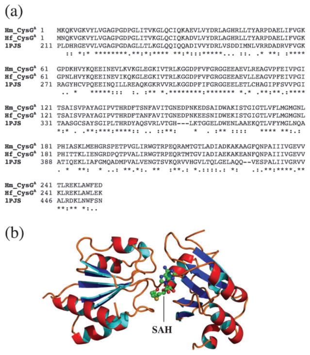
Fig. 4.
(a) Sequence alignment of the CysGA domain of the CysGA–HemD fusion protein from Hb. mobilis and Hp. fasciatum (Hm_CysGA and Hf_CysGA, respectively) with the CysGA domain of the CysG protein of S. enterica for which a crystal structure is available (1PJS). Identical sequence matches in the alignment are indicated by ‘*’, strongly similar matches by ‘:’, and weakly similar matches by ‘.’. (b) 3D model of the CysGA domain for Hb. mobilis based on the above alignment. The position of the bound cofactor SAH (demethylated SAM) is also shown.

