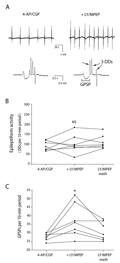
Fig. 1. Co-application of the group I mGluR antagonists LY 367385 (LY) and MPEP did not change the amount of epileptiform activity but did increase the rate of giant GABA-mediated postsynaptic potentials (GPSPs). Slices were exposed first to 4-AP/CGP 55845 and then additionally to LY/MPEP, followed by washout of the LY/MPEP.
A: Extracellular voltage traces recorded from same slice before (left) and after (right) exposure to LY/MPEP. Electrode was placed at stratum lacunosum-moleculare /stratum radiatum border in CA3. Top row: In this example, 6 GPSPs with accompanying discharge deflections (DDs) occurred in a 2-min period before LY/MPEP, and 10 occurred in a 2-min period after LY/MPEP. Bottom row: Single epileptiform events are expanded to reveal a GPSP followed by 4 DDs before LY/MPEP (left) and a GPSP followed by 3 DDs after LY/MPEP (right). The traces in the bottom row were recorded from the same slice as the traces in the top row. B: The epileptiform activity, as measured by total number of DDs per 10-min period, did not consistently change with the addition of LY/MPEP. C: The number of GPSPs per 10-min period increased after addition of LY/MPEP, and decreased with washout of LY/MPEP. * indicates statistically significantly different from control; NS indicates not statistically significantly different from control.

