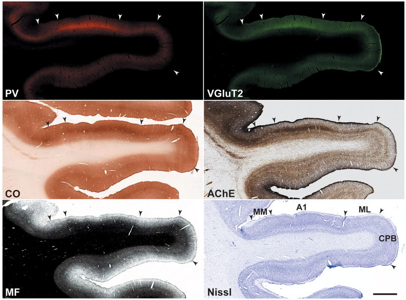
Fig. 2.
Series of adjacent coronal sections through caudal areas of auditory cortex stained for several chemoarchitectonic markers: Parvalbumin (PV), vesicular glutamate transporter 2 (VGluT2); cytochrome oxidase (CO); acetylcholinesterease (AChE); myelinated fibers (MF); Nissl (N). Note that the dense band of staining in layer IIIb/IV is highest in the core area, A1, and weaker in the belt and parabelt areas. Scale bars, 2 mm.

