FIGURE 1.

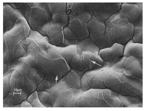
Scanning electron micrograph of bullous keratopathy specimen shows an irregular surface with numerous residual polygonal superficial epithelial cells of varying size and shapes after exfoliation (white arrows).

Scanning electron micrograph of bullous keratopathy specimen shows an irregular surface with numerous residual polygonal superficial epithelial cells of varying size and shapes after exfoliation (white arrows).