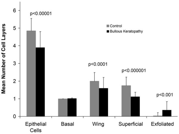
FIGURE 2.

Mean numbers of cell layers in bullous keratopathy versus control specimens. A significant reduction is shown in the total epithelial cell layers and in wing cells and superficial cells in bullous keratopathy specimens. The number of exfoliated cells was significantly increased in bullous keratopathy compared with control specimens.
