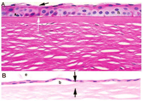
FIGURE 3.

Photomicrographs of epithelial thinning in bullous keratopathy. (A) PAS stain shows thinning of the epithelium with loss of superficial cells (black arrow) and microbullae (white arrow). Hydropic change is evident within the epithelium (h). Many of the wing cell nuclei are faded. (B) Hematoxylin and eosin–stained sections show severe thinning of the epithelium with bullae (b) accompanied by attenuation of the Bowman layer (between black arrows). Exfoliated cells are evident (e). Original magnification, ×250.
