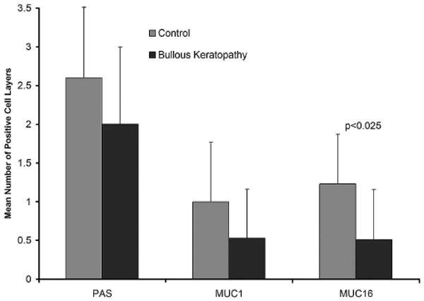
FIGURE 6.

Identification of mucin-positive cell layers in bullous keratopathy compared with control specimens. PAS staining was insignificantly decreased in bullous keratopathy specimens. Immunohistochemical staining for MUC16, but not MUC1, showed a significant decrease in the number of cell layers that were positive.
