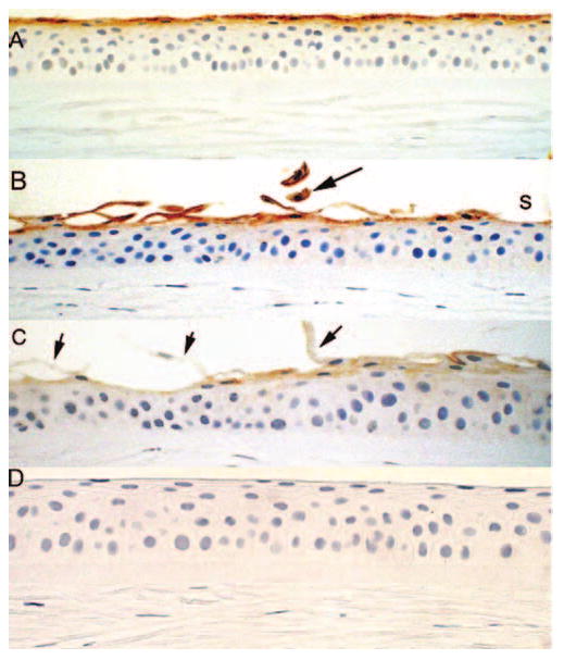
FIGURE 7.

Immunohistochemistry for MUC16 (A) in control specimens shows one to two superficial cell layers staining consistently and (B, C) in bullous keratopathy specimen. (B) Exfoliated cells show intense MUC16 staining (black arrow). Some areas underlying shed cells shows faint staining (s). (C) In some cases exfoliated cells (black arrows) and the underlying epithelium show weak to absent reactivity to MUC16. (D) Negative control of normal cornea with deletion of the primary antibody.
