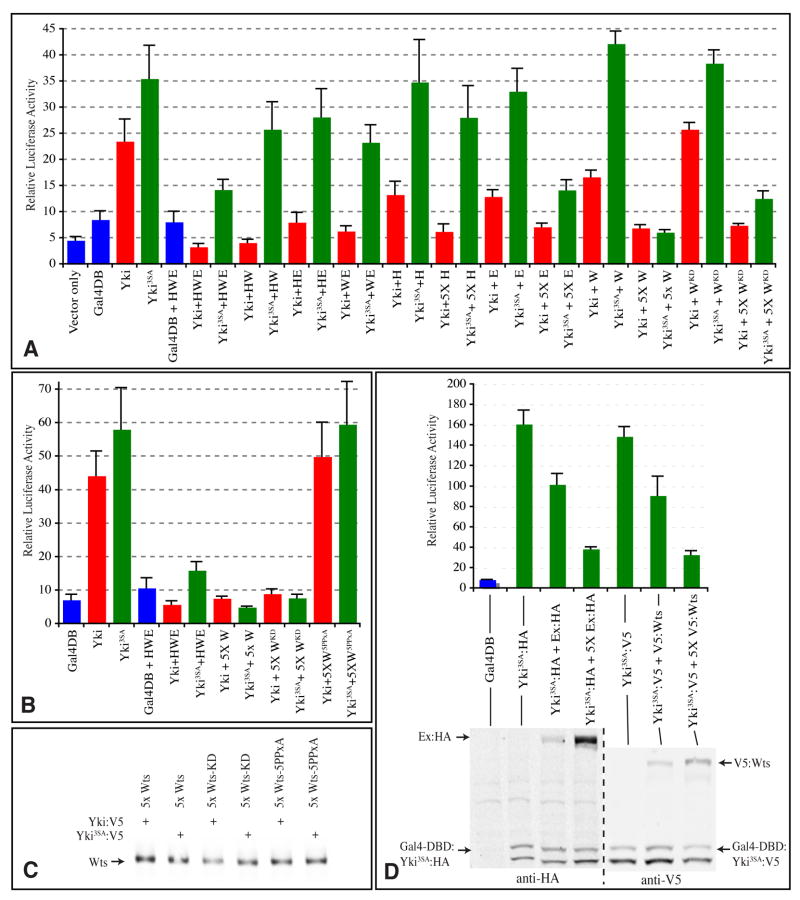
Fig 3. Phosphorylation-independent repression of Yki-mediated transcription.
Expression from a UAS-luciferase reporter is indicated by luciferase activity in lysates of S2 cells transfected to express wild-type (red) or 3SA mutant (green) Gal4-DBD:Yki:V5 fusions (Yki) (or a control Gal4-DBD protein, blue). In A, B, where indicated, Yki transgenes were co-expressed with Flag:Hpo (H), Ex:HA (E), Myc:Wts (W), or Myc:WtsKD or Myc:Wts5xPPxA mutants, using equal amounts of DNA, or, where indicated (5x), a five-fold excess of DNA. Histograms depict the average values from triplicate experiments; error bars indicate standard deviation. The results depicted in A and B are from separate experiments. C) Western blot on lysates from samples in B, showing similar expression of Wts. D) Upper panel shows luciferase activity, lower panel shows Western blot on the same cell lysates. The first three experimental (Yki) samples employed HA-tagged Ex and Gal4-DBD:Yki, and the last three employed V5-tagged Wts and Gal4-DBD:Yki, as indicated. The proteins are distinguished on the Western blots by their mobilities, Gal4-DBD:Yki runs as a doublet.

