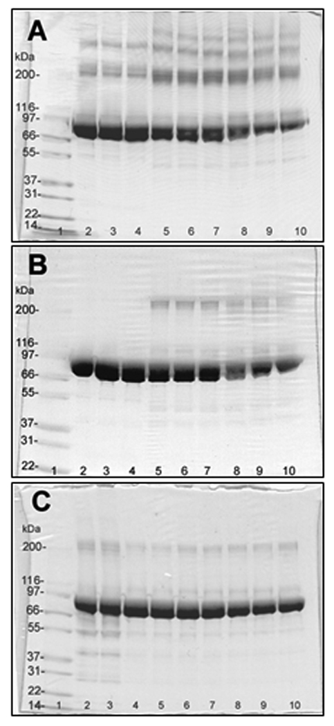
Figure 2.
SDS-PAGE of rabbit muscle phosphorylase b (20 µg/lane) treated with peroxynitrite in the presence of 25 mM NaHCO3 at pH 7.4 (non-reducing conditions). A and B: lanes 2–4 – none; 5–7 – 0.3 mM; 8–10 – 1 mM peroxynitrite. In C: lanes 2 and 3 – reverse-order of peroxynitrite addition (1 mM) and control; lanes 4 and 5 – 0.1 mM; lanes 6 and 7 – 0.3 mM; lanes 8 and 9 – 1 mM peroxynitrite; lane 10 – 1 mM peroxynitrite addition in the absence of NaHCO3 in 25 mM sodium phosphate, pH 7.4. Lane 1 contained molecular weight standard Mark12 (3 µg of each protein). Samples were loaded either 2 hours (A and B) or immediately (C) after treatment with peroxynitrite. In B, samples were reduced with 2 mM DTT at 37°C for 30 min followed by alkylation with iodoacetic acid for 30 min at room temperature in the dark.

