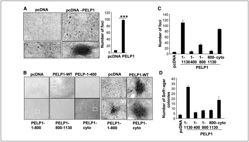
Figure 2.
PELP1 transforms rat kidney epithelial cells (RK3E). A, focus formation in PELP1-transfected or vector-transfected RK3E cells. Right, quantitation of PELP1-induced focus formation. B, RK3E cells were transfected with various domains of PELP1 or a PELP1 mutant that uniquely localizes in the cytoplasm, and focus formation was counted. Representative morphologic characteristics of PELP1-induced and PELP1-mutant induced foci. C, quantitation of PELP1-induced and PELP1 deletion-induced focus formation. D, pooled RK3E transfectants were plated in soft agar, and the colony formations were counted after 21 d. These experiments were repeated thrice; Columns, average of the results.

