Figure 3.
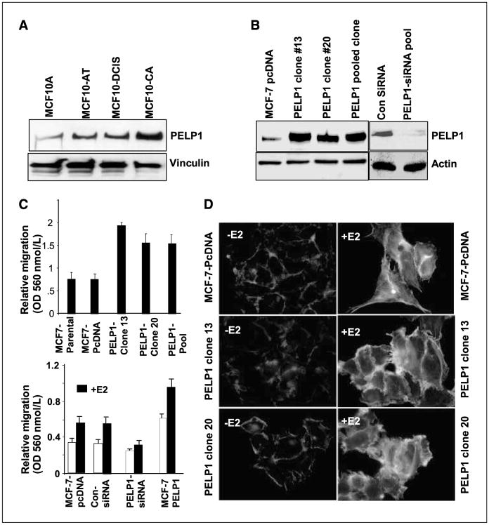
PELP1 promotes cytoskeletal reorganization and motility. A, Western blot analysis of PELP1 expression in lysates from cells derived from the MCF10AT model system. MCF10A, nonmalignant human breast cancer cells; MCF10-AT, weakly tumorigenic; MCF10-DCIS, comedo-type DCIS, highly proliferative, aggressive, and invasive; and MCF10-CA, undifferentiated carcinomas, metastatic. B, total cell lysates from pcDNA- and PELP1-expressing clones (nos. 13, 20, Pool) were analyzed by Western blotting with an anti-PELP1 antibody (left). Actin was used as a loading control. The functionality of PELP1-siRNA was analyzed by Western blotting (right). C, migratory potential of MCF-7 clones overexpressing PELP1 was analyzed by Boyden chamber assay (top). MCF-7 cells stably overexpressing PELP1 and MCF-7 cells transfected with PELP1-siRNA were treated with E2 or were untreated, and the cell migration potential was analyzed by means of the Boyden chamber assay (bottom). D, MCF-7, MCF-7–PELP1 clone nos. 13 and 20 were cultured in a DCC-serum–containing medium, after which they were treated with E2 for 10 min. The status of filamentous actin was visualized by phalloidin staining and was evaluated by fluorescence microscopy.

