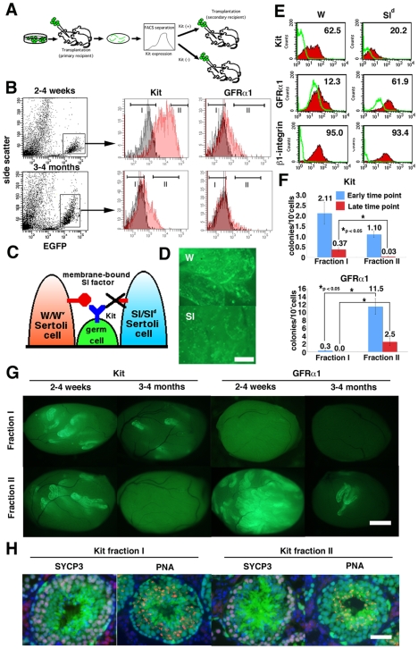
Figure 4. Changes in Kit expression in vivo.
(A) Experimental strategy. After transplantation of GS cells, EGFP-expressing donor cells were fractionated according to Kit or GFRα1 levels. Sorted cells were transplanted into W mice. (B) Fractionation of donor spermatogenic cells. EGFP+ cells were gated and fractionated into two groups according to Kit or GFRα1 levels. Distributions of stained (red) or control (black) are shown. (C) Sl-Kit interaction in W and Sld mice. Germ cells in W mice have a defect in Kit and cannot respond to Sl, whereas Sertoli cells in Sld mice do not express membrane-bound Sl and cannot support differentiation. (D) Appearance of W and Sld recipient testes 2 weeks after transplantation. Differentiation was limited in Sld testis. (E) FACS analysis of W and Sld recipient testis after transplantation. EGFP+ cells were gated for analysis. (F) SSC activity of sorted cells. Both Kit− and GFRα1+ cells showed significant enrichment of SSCs at both time points. (G) Appearance of recipient testes that received sorted cells. (H) Immunohistological section of the recipient testes that received Kit+ or Kit− cells. The donor cells were collected from the primary recipient testes 2 weeks after transplantation, and the recipient testes were stained 2 months after cell sorting. The sections were stained with Rhodamine-PNA (red) for acrosomes and with anti-SYCP3 antibody (blue) for meiotic cells. Bar = 20 µm (D), 100 µm (G), 50 µm (H).

