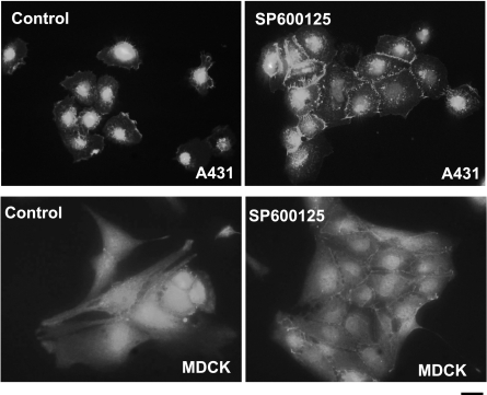
Figure 3.
JNK inhibition leads to cell adhesion in A431 and MDCK cells. Cells were plated in tissue culture-treated glass slides and were then treated with either vehicle (control) or SP600125 (10 μM) for 30 min. After 30 min, the cells were fixed and then stained for E-cadherin. Nuclei were visualized with Hoechst 33258 (solid circles; view ×40). Scale bar = 20 μm.

