Figure 5.
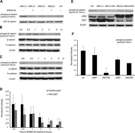
JNK phosphorylates β-catenin. A) In vitro kinase assay using purified activated JNK1α1 or JNK2α2 and GST-β-catenin in the presence or absence of SP600125 (20 μM). Phosphorylated GST-β-catenin was detected using Western blotting, and total GST-β-catenin served as a loading control. This experiment was repeated 5 times with similar results; a representative experiment is shown. B, C) Human keratinocytes (B) and 293T cells (C) were treated with SP600125 for the indicated times, and cell lysates were subjected to Western blotting for phosphor-β-catenin, total β-catenin, and E-cadherin; β-actin served as a loading control. Blot is representative of 3 independent experiments with similar results. D) Lane intensity of phosphor-β-catenin was determined using Kodak gel documentation software and normalized to total β-catenin. Data from 3 independent experiments were plotted as average ± se. E) 293T cells were transfected with plasmids encoding for Flag-JNK1, Flag-JNK2, or the kinase-dead Flag-JNK1DN, and Flag-JNK2DN, and cell lysates were subjected to Western blotting for phosphor-β-catenin and total JNK; β-actin served as loading control. F) Lane intensity of phosphor-β-catenin was normalized to β-actin loading control; data from 3 independent experiments were plotted as average ± se. *P < 0.05 vs. control.

