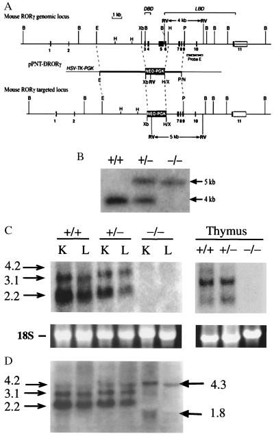
Figure 1.
Targeting the RORγ locus. (A) Schematic representation of the mouse RORγ gene locus, the pPNT-ΔRORγ targeting vector, and the recombination at the RORγ locus. In the targeted locus the region from exon 3 through exon 6 is deleted. DBD, DNA-binding domain; LBD, ligand-binding domain. (B) Diagnostic Southern blot analysis. Genomic DNA was cut by EcoRV, electrophoresed, and hybridized to the 3′-flanking probe E (indicated in A), which detected fragments of the expected size of 4.0 kb for wild-type and 5.0 kb for the mutant allele. Lanes indicate digested DNA from wild-type (wt; +/+), heterozygous (+/−), and homozygous mutant (−/−) mice. (C and D) Northern blot analysis was performed with RNA isolated from liver (L), kidney (K), and thymus of wt, RORγ+/−, and RORγ−/− mice by using either a probe encompassing the deleted region encoded by exons 3–6 (C) or a full-length RORγ probe (D).

