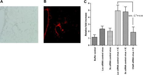
Figure 1.
Sections of mouse lung after intratracheal administration of adenovirus and induction of IgGIC-induced injury. A, B) Assessment of adenovirus distribution in lung after intratracheal administration of dsRed fluorescence protein-expressing adenovirus. Four days after virus injection, lung sections were examined, and dsRed protein was visualized in the lissamine-rhodamine channel under fluorescence microscopy. A) Bright-field image of infected lung (×20). B) Abundant red fluorescence of lung receiving dsRed virus in bronchiolar (left) and alveolar (center) walls (×20). C) Four days after intratracheal injection of either buffer control (PBS), C5aR-siRNA silencing virus (C5aR-siRNA), luciferase control virus (Luc siRNA control virus), or scrambled siRNA control virus (Sc siRNA control virus), mice were subjected to IgGIC-induced lung injury. Lung total RNA was isolated 4 h after injection of IgGIC injury and analyzed for mRNA for C5aR by real-time PCR analysis. Data are expressed as relative fold changes over nonvirus infected, control lungs (buffer control group). Results are means ± se; ≥5 mice/group.

