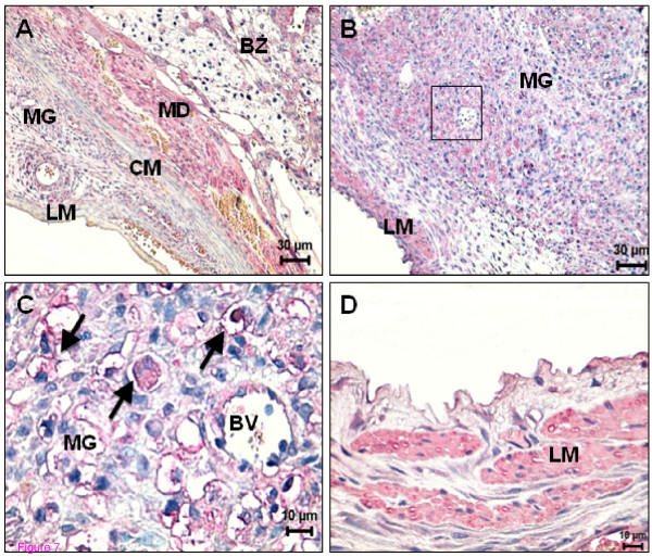
Figure 7.
Spatial distribution of CB1 and TRPV1 in the uterus on day 16 and 19 of pregnancy. (A) On day 16, decidual and trophoblast cells from the basal zone are positive for CB1. Note the lack of expression in circular and longitudinal muscle layer; (B) In the metrial gland, the expression of TRPV1 at day 19 is mainly localized in uNK cells and in longitudinal muscle layer. (C) High power of the square depicted in (B). Expression of TRPV1 is particularly evident in uNK cells (arrows). (D) High power showing an intense positive signal for TRPV1 in the longitudinal muscle layer. BV - Blood vessel; BZ - Basal zone; CM- Circular muscle layer; LM - Longitudinal muscle layer; MD - Mesometrial decidua; MG - Metrial gland.

