Fig. 1.
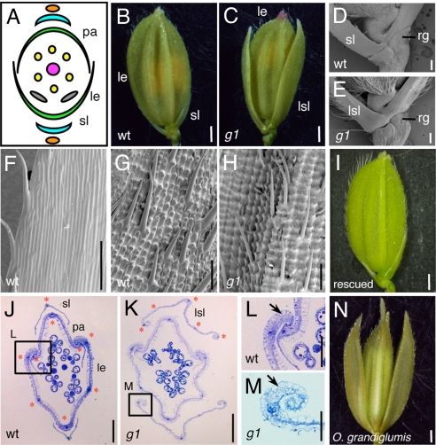
Spikelet phenotypes of the g1 mutant. (A) Schematic representation of the spikelet of WT rice. (B) A WT spikelet. (C) A g1 spikelet showing the LSL. Basal region of the spikelet in WT (D) and g1 (E). Epidermal surface of the sterile lemma (F) and the lemma in WT (G). (H) Epidermal surface of the LSL in g1. (I) A spikelet in a rescued g1 mutant, which was transformed with a genomic fragment encompassing the G1 locus. Cross section of the spikelet in WT (J) and g1 (K). Close-up view of the marginal region of the WT lemma (L) and g1 LSL (M). Red stars indicate vascular bundles (J and K). Arrows indicate the peripheral structure of the lemma and LSL (L and M). (N) A spikelet of O. grandiglumis (W0613). le, lemma; pa, palea; rg, rudimentary glume; sl, sterile lemma. (Scale bars: 1 mm in B, C, I, and N; 200 μm in J and K; 100 μm in D, E, F, G, and H; and 50 μm in L and M.).

