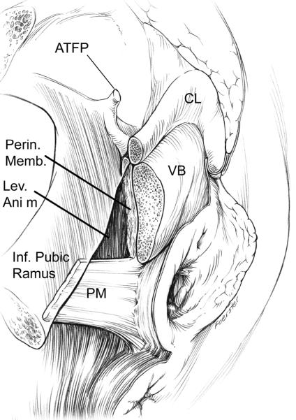
Figure 1.
Drawing of dissection revealing the perineal membrane (PM) showing its lateral attachment to the inferior pubic ramus. A window in the perineal membrane has been cut to reveal the attachment of the levator ani muscle (LA) and its fusion with the vestibular bulb (VB). Extension to the arcus tendineus fascia pelvic is also shown (ATFP) which is shown inside the pubic bone attaching to its inner surface; clitoris (CL). (© DeLancey 2007)

