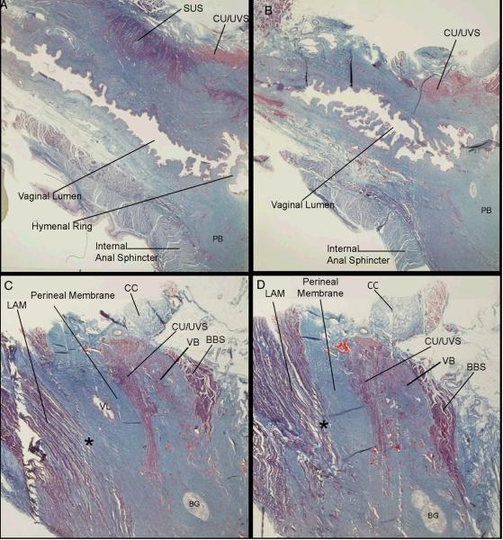
Figure 4.
Histologic (trichrome) sections of an adult female cadaver in sagittal plane beginning in the parasagittal plane just lateral to the urethral lumen (A) and progressing laterally to the region of the perineal membrane demonstrating the insertion of levator ani muscle (LAM) fibers directly into the cranial portion of the perineal membrane (C, D) lateral to the vagina (*). In this region, the perineal membrane can be seen to be fused with surrounding structures. Abbreviations: bulbospongiosus muscle (BBS); Bartholin's gland (BG); clitoral crus (CC); compressor urethrae/urethro-vaginal sphincter muscles (CU/UVS); levator ani muscles (LAM); perineal body (PB); striated urethral sphincter (SUS); vaginal lumen (VL); vestibular bulb (VB). (© DeLancey 2007)

