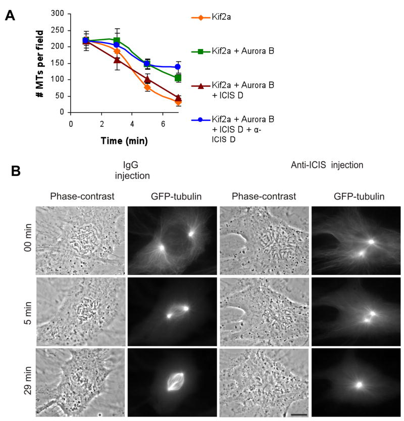
Figure 2. Injection of α-ICIS antibodies (AB) into cells gives a monopolar spindle phenotyope.
A. ICIS D antibodies inhibit Kif2a reactivation in vitro. Quantification of the mean number of microtubules per field in the presence of recombinant Kif2a, Aurora B, ICIS D, and α-ICIS. Error bars represent standard deviation.
B. IgG control-injected and α-ICIS injected in prophase Xenopus S3 cells expressing GFP-tubulin. Time lapse images from Supplemental movies 1 and 2. 0 minutes=time of injection. Scale bar represents 10μm.

