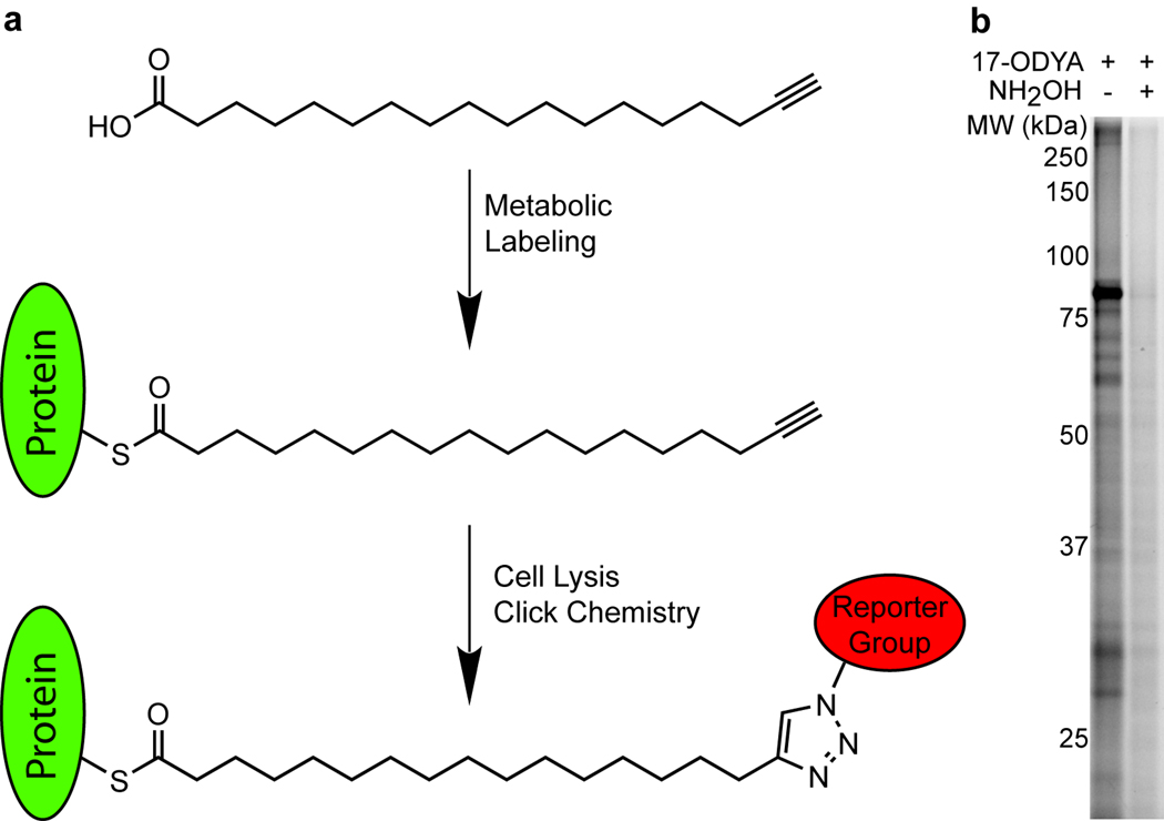
Figure 1.
17-ODYA labeling and detection of palmitoylated proteins. (a) Schematic of 17-ODYA labeling. Cultured Jurkat T-cells were metabolically labeled with 17-ODYA and the lysates were then reacted with rhodamine-azide or biotin-azide for gel-based and LC-MS-based characterization of palmitoylated proteins. (b) Profiling palmitoylated proteins in the membrane fraction of Jurkat T-cells incubated with 25 µM 17-ODYA for 8 hours. Half of the sample was boiled in 2.5% hydroxylamine (NH2OH) for 5 minutes to hydrolyze thioesters and remove 17-ODYA labeling.

