
Figure 2.
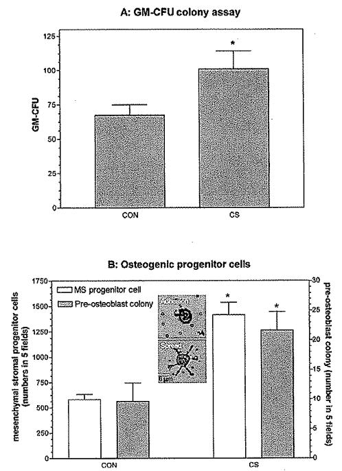
CS effects on BM cells in vitro. (A) CS enhanced the survival of GM-CFUs from BM cells. BM cells were harvested from the tibia and femur and cultured in RPMI medium for one day. Nonadherent BM cells were then taken and cultured with or without CS (500 μg/ml) in premixed methylcellulose culture medium containing IL-3 and GM-CSF. GM-CFUs were counted on day 7. (B) CS enhanced the survival and clonogenicity of MSCs. BM cells were seeded in 12-well culture plates (3 × 10³/well) with or without CS (500 μg/ml). After one day, the number of adherent MSCs identified by their pseudopodia processes (see the insert) that were left on the culture plate after removal of nonadherent cells was scored in five fields by microscopy (magnification: ×100). In addition, the number of preosteoblast colonies that developed from these cells was scored on day 5 in five fields by microscopy (magnification: ×100). Values are the means ± 1 SEM for six determinations per group. Data and error bars represent the means ± 1 SEM for one representative of three experiments, as analyzed by GraphPad Prism software version 3.03. *, P < 0.01 by Student’s t test (compared with control).