
Fig. 1.
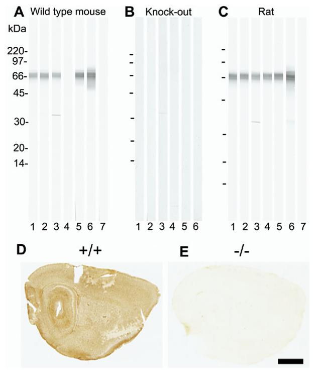
Specificity testing of GLT/EAAT2 antibodies by immunoblotting (A—C) and by immunocytochemistry (D—E). Whole brain tissue from wild-type (+/+) mouse (A), EAAT2 knockout (−/−) mouse (B) or adult Wistar rat (C) was solubilized in SDS, run on SDS-PAGE (10–20% gradient gels; 250 μg protein per gel) and blotted onto nitrocellulose. The blots were cut into strips (25 strips per blot) and labeled with the following antibodies: 1: 0.2 μg/ml anti-B12 (AB#360); 2: 0.2 μg/ml anti-B12 (AB#150), 3: 0.1 μg/ml anti-B493 (AB#95), 4: 0.5 μg/ml anti-B518 (AB#94), 5: 0.2 μg/ml anti-B563 (AB#355), 6: 0.1 μg/ml anti-73 kDa (AB#171) and 7: no primary antibody (negative control). Mice brains (D: +/+;E: −/−) were immersion fixed in 4% formaldehyde. The sections were incubated with anti-EAAT2 antibodies in the presence of Triton X-100, followed by appropriate biotinylated secondary antibodies, then streptavidin peroxidase, and finally developed with diaminobenzidine. The figure only shows the test results for one of the antibodies: anti-B563 (2 μg/ml; AB#355). Scale bar=1.5 mm.