
Fig. 6.
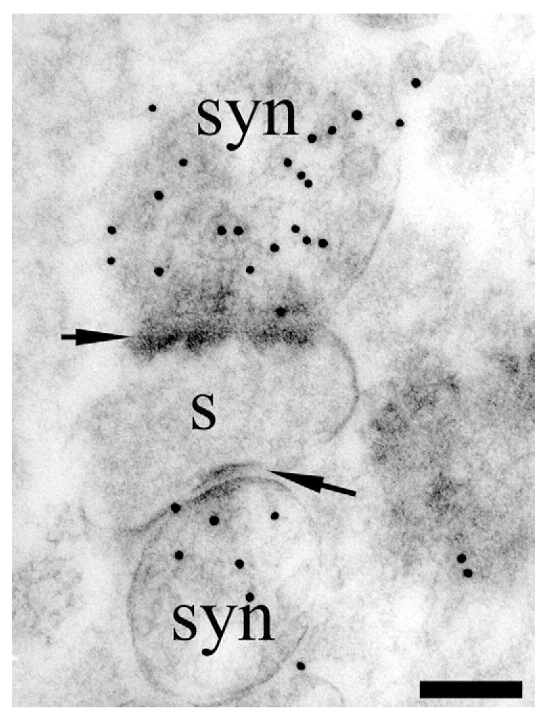
TEM visualization of D-aspartate uptake in synaptosome preparations. Rat synaptosomes (syn) were incubated for 20 min in 50 μM D-aspartate. Note heavy labeling over synaptosomes (syn) that are attached to a remnant of a dendritic spine (s). The synaptosomes are clearly identifiable by the presence of synaptic densities in the opposing membranes (arrows) and vesicles. Antibody: 482 D-Asp. Scale bar=100 nm.