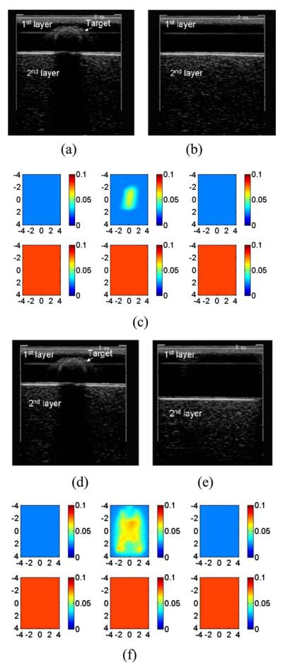
Fig. 5.
Reconstructed absorption map of a 1-cm-diam spherical target with calibrated optical properties μa=0.07 and , located at (x,y,z)=(0,0,0.9 cm). (a) B-scan ultrasound image of a two-layer medium with the target. A plastisol phantom was located at 1.4 cm underneath the probe surface with a zero-degree tilting angle. (b) B-scan ultrasound image of the phantom without the target. (c) Reconstructed target absorption map at 780 nm. (d) B-scan ultrasound image of the two-layer medium with the target [same as (a)]. (e) The two-layer medium with the plastisol phantom located at 2 cm with a zero-degree tilting angle. (f) Reconstructed target absorption map at 780 nm.

