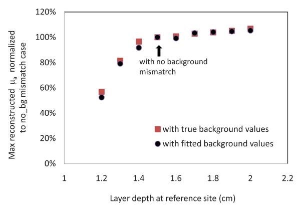
Fig. 6.
Percentage of the maximum reconstructed μa normalized to the no-background mismatch case as a function of the two-layer interface depth at the reference site (phantom data) using both calibrated background and fitted background optical properties values. The two-layer interface at the target site is located at 1.5 cm depth from the probe.

