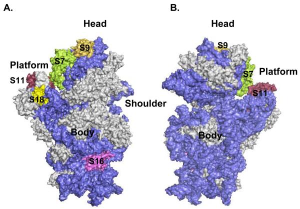
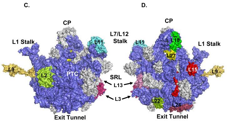
Figure 4.
3D-Models of the E. coli ribosomal subunits displaying the location of phosphorylated mitochondrial ribosomal proteins. The phosphorylated ribosomal proteins are highlighted in different colors, while gray represents unphosphorylated ribosomal proteins, and blue is the rRNA. Coordinates of the E. coli 30S subunit and 50S subunit were obtained from the Protein Data Bank (Acc. # 2AW7 and 2AW4). (A) The 30S subunit is illustrated from the solvent side, while (B) represents a view of the small subunit from the 50S interface. (C) Representation of the 50S subunit from the 30S interface, while (D) is a view of the large subunit from the solvent side. Specific regions, peptidyl transferase center (PTC), central protuberance (CP), sarcin-ricin loop (SRL), L1 and L7/L12 stalks, exit tunnel, and the phosphorylated ribosomal proteins were labeled in the model generated by PyMOL software.


