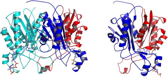
Figure 1.
Ribbon diagrams of the PqsD monomer and dimer. In the dimer representation one monomer is shown in cyan and the other is colored by domain with residues 1-174 in blue and 175-329 in red. Anthranilate-modified Cys112 and anthraniloyl-CoA molecules are shown as sticks and illustrate the locations of the active sites and CoA binding tunnels. The PqsD monomer is colored by domain as in the dimer and is rotated slightly to illustrate the structural similarity between the two domains.

