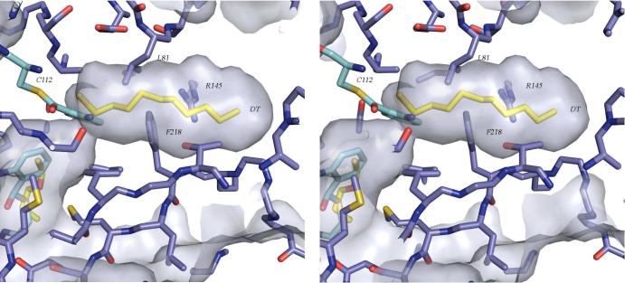
Figure 7.
Stereoview of the presumed acyl binding pocket of PqsD illustrating that a conformational change is likely required in order to accommodate β-ketodecanoate. Decanethiol from the MtFabH structure (PDB code 2QO1) was superimposed as in Figure 7 and is shown with yellow carbon atoms. In the observed conformation, Phe218, Leu81, and Arg145 are among the residues limiting access to the binding pocket.

