
Figure 1.
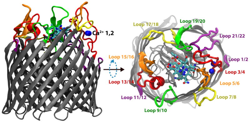
Membrane-plane and extracellular views of the Ca2+/CN-Cbl-bound structure of BtuB. The barrel and luminal domains are colored in grey, the two Ca2+ ions are in blue, and CN-Cbl is shown in a stick representation colored by atom type. The eleven extracellular loops are drawn using a thicker representation and labeled using different colors to distinguish them from their neighbors.