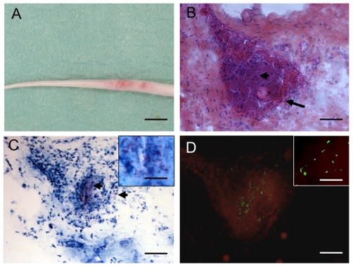
Figure 3. Histological study of serial mouse tail sections following mouse-tail infection with M. ulcerans-GFP.
(A) Fifty days after subcutaneous inoculation in the tail of 105 bacilli showing the beginning of ulceration. (B) Hematoxylin phloxine saffron staining on tissue section reveals massive necrosis. (C) Ziehl-Nieelsen staining showing many extracellular individual and clustered bacilli (arrowheads) associated with necrosis, and (D) bacilli expressing GFP. Scale bars: 0.8 cm (A), 100 µm (B, C, and D) and 40 µm (C and D insets).

