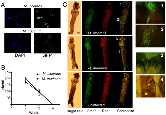
Figure 4. Concentration of M. ulcerans-GFP within 4th instar mosquito larvae.
(A) Epifluorescence microscopy (x100 objective) demonstrating that the majority of total fluorescent bacteria (DAPI-staining, blue filter) are also GFP-expressing M. ulcerans (JKD8083) and M. marinum (JKD8062) (green filter). (B) Reduction of M. marinum and M. ulcerans within the water of the microcosms over the 4-week duration of the experiment as measured by qPCR. Results are the means and standard deviations of four biological repeats. (C) Bright field, red fluorescence, green fluorescence and composite images showing the accumulation of M. ulcerans around the mouth and mid-gut of a 4th instar Aedes camptorhynchus larva. Scale bar indicates 0.5 mm. Inset images are ×400 magnification. No specific green fluorescence was observed in larvae infected with M. marinum or uninfected controls. Red fluorescence images were included to show that green fluorescence was due to GFP expression and not autofluorescence.

