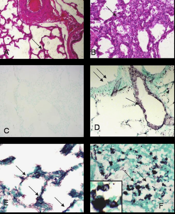
Figure 3.
Histopathology and NMT1 expression: lung sections from control calves (A) showed normal tissue architecture (arrow) while those from the infected calves (B) showed extensive inflammation (arrow). Lung sections stained with only secondary antibody (C) did not show any staining while von Willebrand Factor antibody (D) reacted with vascular endothelium (single arrow) but not bronchiolar epithelium (double arrows). Lung sections from control calves (E) showed light staining for NMT in alveolar septa but the staining was intense in neutrophils (F) in inflamed lung. Note NMT staining nuclei and cytoplasm of neutrophils (inset). Original magnification × 100. (A color version of this figure is available at www.vetres.org.)

