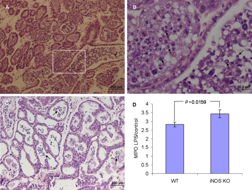
Figure 6.
Neutrophil recruitment into milk spaces 24 h after intramammary infusion of 10 μg LPS in iNOS −/− (A and inset of A inB) and wt C57BL/6 mice (C). H&E staining of formalin-fixed mammary tissues (A−C). Neutrophil recruitment to the mammary gland was assessed by the MPO assay (D). Significant difference in relative (LPS to PBS-challenged gland) MPO activity was observed in the mammary glands of iNOS −/− mice compared with C57BL/6 mice. Data are mean and S.E., n ≥ 6/group and p values represent comparison between groups using the Student t-test. Scale bars 200 μm (A and C), 50 μm (B). (A color version of this figure is available at www.vetres.org.)

