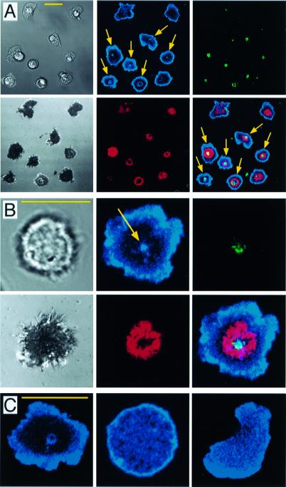
Figure 1.
Localization of CD45 to the central region of the mature immunological synapse. I3/2.3-Alexa546-labeled T cells were settled onto lipid bilayers containing Cy-5-conjugated ICAM-1, and Oregon green-conjugated agonist MHC-peptide. ICAM-1 (red), MHC-peptide (green), CD45 (blue). (Scale bars = 10 μm.) (A and B) Interference reflection microscopy and fluorescent images of the T cell/bilayer interface. (Upper Left) Transmitted light. (Lower Left) Interference reflection microscopy. (Lower Right) Red/green/blue overlay. n = 300. (C) Fluorescent images of CD45 density at the cell/bilayer interface. (Left) Peptide/MHC/ICAM substrate. (Middle) Poly-l-lysine substrate. (Right) ICAM-1 substrate.

