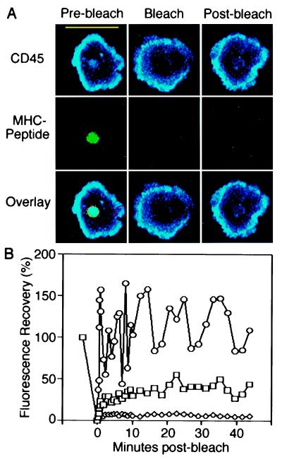
Figure 5.
Photobleaching recovery of CD45 within the immunological synapse. (A) T cells labeled with I3/2.3-Cy5 Fab fragments were allowed to form the mature immunological synapse on planar bilayers containing unlabeled ICAM-1 and Oregon green-conjugated MHC-peptide. Fluorescence from accumulated MHC-peptide (green) and CD45 (blue) was quenched by photobleaching at the position shown (red circles). (Scale bar = 10 μm.) (B) Bleaching recovery kinetics are represented as the percentage recovery of fluorescence after photobleaching for CD45 (open squares) and MHC-peptide (open diamonds) within the immunological synapse, and also for CD45 at the periphery of the cell/bilayer contact region (open circles). Data are representative of five individual experiments.

