
Figure 2.
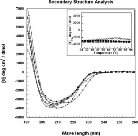
CD spectra of VrD1 and its mutants. The secondary structure was analyzed by circular dichroism spectroscopy. The recombinant VrD1 (50 μM) was dissolved in 20 mM phosphate buffer, and the CD spectrum was scanned from 190 to 260 nm. The CD signals in millidegree were converted to mean-residue ellipticity [θ]. The inset shows temperature melting curves of the VrD1 and VrD1 mutants. Closed quadrate: wild type, open circle: G9A, open quadrate: W10A, open diamond: T17A, open triangle: K24A, and cross: C3AC46A double mutant.