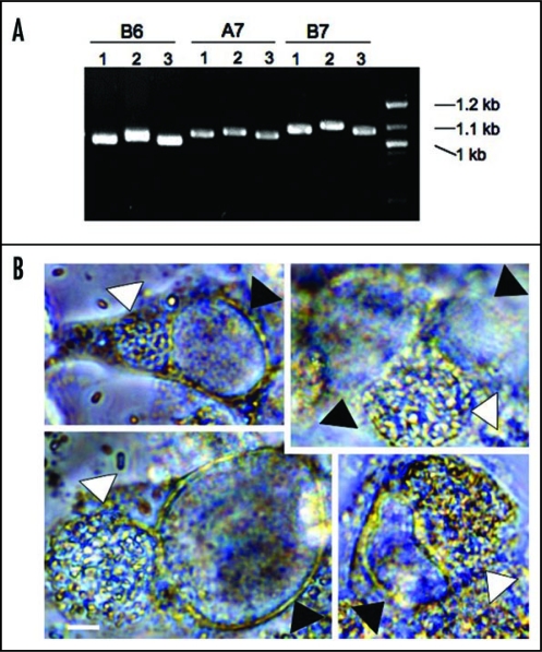
Figure 3.
Genetic variation of tandem repeat DNAs in different E. cuniculi isolates and co-infection of E. cuniculi and C. trachomatis. (A) Examples of TR DNAs. Three E. cuniculi isolates (1 is for EC1, 2 for EC2 and 3 for EC3) have different sized banding patterns. B6, A7 and B7 are TR areas chosen with the Tandem Repeat Finder (see the text). (B) E. cuniculi and C. trachomatis infect the same host cell forming two independent parasitophorous vacuoles. White arrowheads indicate E. cuniculi vacuoles and black arrowheads indicate C. trachomatis vacuoles. Scale = 5 µm.

