Fig. 1.
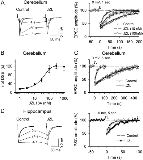
MAGL inhibitor JZL184 (JZL) potentiates DSE mouse cerebellar Purkinje neurons and DSI in mouse hippocampal pyramidal neurons. A, Left, examples of EPSCs in Purkinje neurons 4 s before (−4 s), 4 s, and 60 s after the depolarization (1 s from −60 to 0 mV), in the presence of the solvent (DMSO, control) or JZL184. Right, time course of averaged DSE in slices treated with DMSO (control, n = 8) and different concentrations of JZL184 (JZL, 10 nM, n = 8; 100 nM, n = 6). The lines are single exponential fitting curves of the decay of DSE. B, JZL184 caused a dose-dependent increase in the decay time constant (τ) of DSE (n = 4–8 for each point). C, JZL184 (1 μM) dramatically prolonged DSE when longer depolarization (5 s from −60 to 0 mV) was used to induce DSE (n = 6–8). D, Left, examples of IPSCs in hippocampal pyramidal neurons 4 s before (−4 s), 0 s, and 24 s after the depolarization (5 s from −60 to 0 mV), in the presence of the solvent (DMSO) or JZL184. Right, time course of averaged DSI in slices treated with DMSO (control, n = 10) and 100 nM JZL184 (n = 9).

