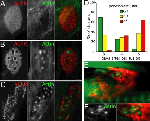
Fig. 1.
Actin–rich puncta lie within the perforations of AChR aggregates. C2C12 myotubes cultured on laminin were incubated with BTX (red) and phalloidin (green) to label AChR and F-actin, respectively. (A) In immature AChR plaques (day 3 after cell fusion) fine actin cables lie below aggregates. (B) As plaques become perforated (day 4 after cell fusion), actin-rich puncta appear within perforations. (C) As aggregates become branched (day 5 after cell fusion), they bear multiple actin-rich puncta. (D) The number of actin-rich puncta increases during maturation of the AChR clusters in C2C12 myotubes (n = >45 per time point). (E) Similar actin-rich structures were found in perforations at the developing NMJ (postnatal day 8). Tibialis anterior muscle was infected with adenovirus expressing β–actin-GFP (green) and counterstained with BTX (red). Area bracketed in (E) is shown in higher magnification in (F). (Scale bars, 5 μm.)

