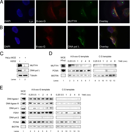
Fig. 1.
Recruitment of BER proteins to the sites of oxidative DNA damage. The experiments were done under the conditions as specified in Materials and Methods. (A and B) Fluorescent microscope images of 355-nm laser-irradiated HeLa cells synchronized in G1/S phase and stained with antibodies against 8-oxo-G and MUTYH (A) or DNA pol λ (B). Colocalization was observed in 50% of the analyzed cells. (C) Immunoblot analysis of MUTYH and DNA pol λ protein level, 5 h upon recovery, in HeLa whole cell extract (WCE) treated with and without H2O2. (D and E) Protein recruitment assay using A:8-oxo-G (Left) or C:G (Right) biotinylated hairpin substrate and HeLa WCE in the absence (D) or presence (E) of Mg2+ for the times indicated (*, the higher molecular weight band corresponds to MUTYH localized in mitochondria). Only proteins that bind to hairpin substrate can be cross-linked and visualized by immunoblotting.

