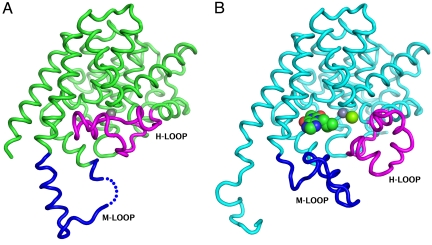
Fig. 4.
Crystal structures of the isolated catalytic domains also show “open” and “closed” conformations of the H-loop. The Cα trace of the isolated catalytic domain (579–919) is shown as green tubes. The H-loop (700–723) and M-loop (830–856) are colored magenta and blue, respectively. (A) Structure of the unliganded catalytic domain that shows the H-loop folded into the catalytic site and displacing the Mg2+ ion. The Zn2+ ion is shown as a gray sphere. (B) Structure of the catalytic domain co-crystallized with IBMX, showing the H-loop swung out. Zn2+ and Mg2+ ions, shown as gray and green spheres, are both visible in this structure.

