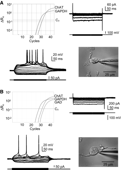
Fig. 6.
Properties of ChAT+/GAD+ neurons resemble those of ChAT+ neurons rather than GAD+ neurons. BF neurons in which ChAT sequence is detected by RT-PCR resemble one another and stereotypical BF cholinergic neurons regardless of the detection of GAD sequence. Each example shows the real-time PCR amplification plot (top left), voltage-clamp records of the inward rectifier current in response to hyperpolarizing voltage steps from a holding potential of −60 mV (top right), current-clamp data of superimposed membrane voltage records during a series of small positive and negative current steps from −60 mV (bottom left), and a photo of the acutely dissociated neuron (bottom right). A: ChAT+ neuron. B: ChAT+/GAD+ neuron. Both examples are from aged rats. Current-clamp solutions (see methods) were used for this set of experiments. ChAT, choline acetyltransferase; GAPDH, glyceraldehyde-3-phosphate dehydrogenase; GAD, glutamic acid decarboxylase; CT, detection threshold; ΔRn, change in reporter fluorescence.

