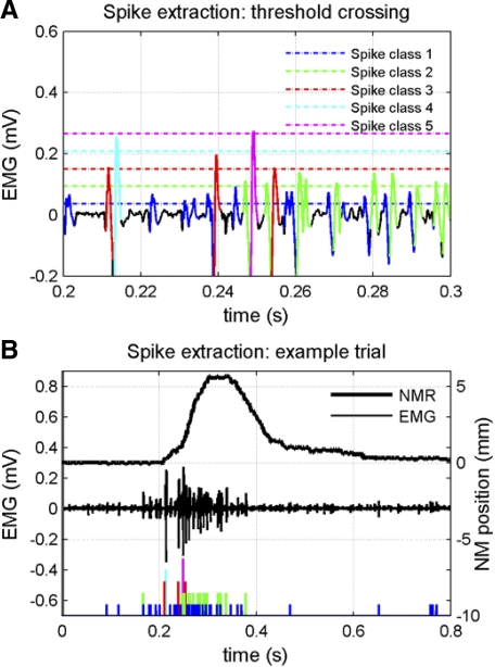
Fig. 1.
Example of spike extraction and classification for electromyogram (EMG) of retractor bulbi muscle (subject RB4). A: a portion of EMG record shown with expanded time base to illustrate threshold crossing criterion for spike extraction and classification for 5 spike amplitude classes. B: entire EMG record for a conditioned-stimulus alone trial (middle trace), showing extracted and classified spikes (bottom trace) and conditioned nictitating membrane response (NMR; top trace). The record is the same as that used in Fig. 1 of Lepora et al. (2007), with A shifted by −50 ms to show the tallest spikes. (The threshold was erroneously shown at too high a value in that figure, although the correct value of 0.0375 mV was used correctly elsewhere in the study.)

