
Figure 4.
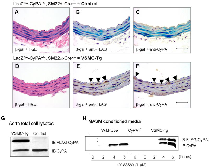
Characterization of CyPA expression in VSMC-Tg mice. (A–F) Representative immunostaining and β-gal staining of aorta from VSMC-Tg mice (LacZflox-CyPA+/SM22α-Cre+) and control mice (LacZflox-CyPA+/SM22α-Cre−). β-gal staining shows >90% expression only in VSMC (B) and complete excision with SM22α-Cre (E). FLAG-CyPA was expressed only in this VSMC-Tg mice but not in control mice. There was no change in endogenous CyPA expression. Scale bars, 50 μm. (G) Western blot demonstrated that FLAG-CyPA was expressed only in the VSMC-Tg mice aorta, but not in control mice. (H) Mouse aortic VSMC were stimulated with 1 μM LY83583 for the indicated times, conditioned media prepared, and secreted CyPA and FLAG-CyPA were detected by western blotting.