
Figure 10.
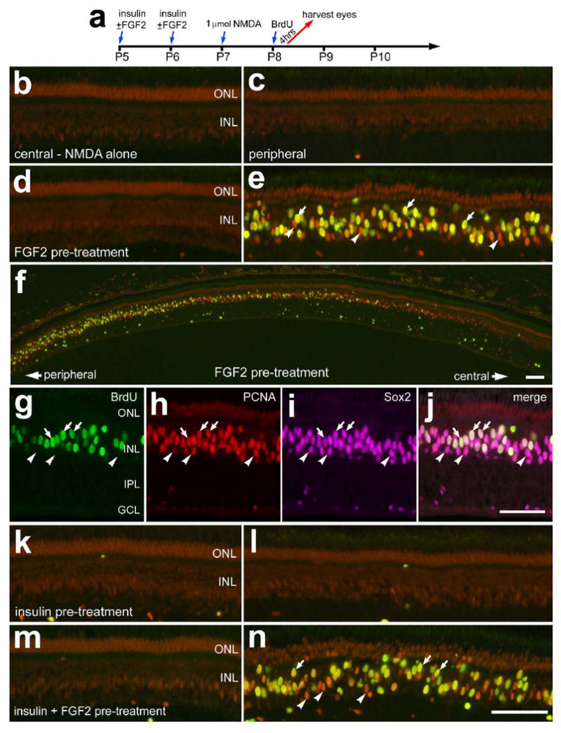
The proliferation of Müller glia in damaged retinas is potentiated by FGF2, but not insulin, prior to acute damage. Retinas were obtained from eyes that received 2 consecutive daily injections of vehicle or growth factor at P5 and P6, 1 μmol of NMDA at P7, 2 μg of BrdU at P8 and harvested 4hrs after the final injection (a). Eyes were injected with vehicle (b and c), FGF2 alone (d-j), insulin alone (k and l), or the combination of insulin and FGF2 (m and n). Vertical sections of the retina were labeled with antibodies to PCNA (red), BrdU (green), and Sox2 (magenta). Arrows indicate the nuclei of Müller glia that were immunoreactive for BrdU and PCNA (e and f) or BrdU, PCNA and Sox2 (g-j). Arrow-heads indicate the nuclei of Müller glia that are labeled for PCNA alone (e and f) or PCNA and Sox2 (g-j). The calibration bar (50 μm) in panel f applies to panel f alone, the bar in j applies to g-j, and the bar in n applies to a-e and k-n. Abbreviations: ONL – outer nuclear layer, INL – inner nuclear layer, IPL – inner plexiform layer, GCL – ganglion cell layer.