
Figure 2.
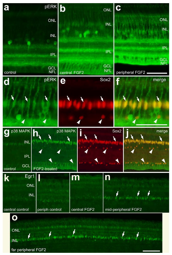
Short-term exposure to FGF2 stimulates the accumulation of pERK, p38 MAPK and Egr1 in Müller glia in peripheral regions of the retina. Vertical sections of the retina were labeled with antibodies to pERK (green; a-d and f), Sox2 (red; e, f, i and j), p38 MAPK (green; g, h and j) or Egr1 (k-o). Arrows indicate the nuclei of Müller glia that are labeled for pERK and Sox2 (d-f), p38 MAPK and Sox2 (h-j), or Egr1 (k and o). The arrow-heads indicate Sox2-positive nuclei of cholinergic amacrine cells in the INL and displaced to the GCL (d-f and h-j). The calibration bar (50 μm) in panel c applies to a-c, the bar in f applies to d-f, and the bar in o applies to g-o. Abbreviations: ONL – outer nuclear layer, INL – inner nuclear layer, IPL – inner plexiform layer, GCL – ganglion cell layer.