
Figure 4.
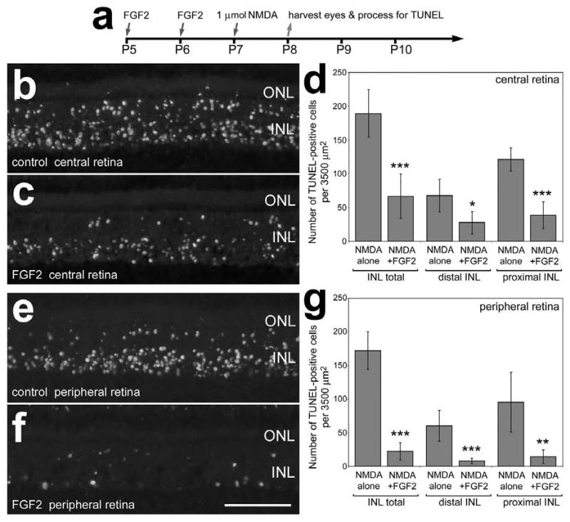
Intraocular injections of FGF2 prior to NMDA-induced retinal damage results in less cell death. Retinas were obtained from eyes that were injected with 300 ng FGF2 at P5 and P6, 1 μmol of NMDA at P7, and harvested 24 hours later at P8 (a). Vertical sections of central (b and c) and peripheral (e and f) regions of the retina were labeled using the TUNEL method. Panels d and g are histograms illustrating the means and standard deviations for numbers of TUNEL-positive cells in distal (presumptive bipolar and horizontal cells) and proximal (presumptive amacrine cells) in central (d) and peripheral (g) regions of the retina. Significance (*p<0.01, **p<.001, ***p<0.0001) of difference was determined by used a two-tailed student's t-test. The calibration bar (50 μm) in panel f applies to panels b, c, e and f. Abbreviations: ONL – outer nuclear layer, INL – inner nuclear layer.